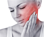
Understanding Trigeminal Neuralgia
Trigeminal Neuralgia
Anatomy of trigeminal nerve
The trigeminal nerve is the largest of 12 cranial nerves. Its main function is transmitting sensory information to the skin, sinuses and mucous membranes in the face. It also stimulates movement in the jaw muscles.
The trigemnial nerve has three different divisions. Each division has different functions .

Opthalmic division :
The opthalmic division conveys sensory information from the :- scalp
- forehead
- upper parts of the sinuses
- upper eyelid and associated mucous membranes
- cornea of the eye
- bridge of the nose
Maxillary division :
Like the opthalmic division, the maxillary division of trigemnial nerve has a sensory component. It transmits sensory information from the :
- lower eyelid and associated mucous membranes
- middle part of the sinuses
- nasal cavity and middle part of the nose
- cheeks
- upper lip
- some of the teeth of the upper jaw and associated mucous membranes
- roof of the mouth
Mandibular division :
The mandibular division is the only part of trigeminal nerve that has both sensory and motor functions . It communicates sensory information from the :
- outer part of the ear
- lower part of the mouth and the associated mucous membranes
- front and middle parts of the tongue
- teeth of the lower jaw and the associated mucous membranes
- lower lip
- chin
It also stimulates movement of the muscles in the jaw and some of the muscles within the inner ear .
Definition of Trigeminal Neuralgia:
It is a condition of the 5th cranial nerve, trigeminal nerve which is characterized by paroxysms of sudden intense pain in distribution of one or more branches .It can be described as stabbing, shooting or electric shock like pain in face which lasts for a minute at a time. The pain ends as abruptly as it starts. Paroxysms can occur with any stimulation of the terminals of affected nerve branches such as washing face, shaving, brushing teeth and drinking.
Types :
Typical / Classical :
(previously termed classical, idiopathic and essential trigeminal neuralgia ) With typical trigeminal neuralgia, the most common form, patients suffer from unpredictable episodes of stabbing, electric shock like pain in a consistent location. Usually caused by the blood vessel compressing the trigeminal nerve root. The pain can be reproduced by touching a "trigger point" on the face or performing a certain activity, such as chewing or talking.Atypical / Symptomatic :
Patients with atypical trigeminal neuralgia experience a persistent dull ache or burning sensation in one part of the face. However episodes of sharp pain can point for the pain and it can grow worse over time.
Causes :
The condition has no clear cut cause. Some of the major theories that may cause trigeminal neuralgia are :
Typical neuralgia :
- Superior cerebellar artery / Tortuous vein compression : The artery may loop on the nerve and compress the trigeminal nerve root when the blood vessel is repeatedly rubbing on the nerve. It causes losing of myelin sheath and causes demyelination of fast conducting fivers at multiple points. They are diseased and hyper excited. It activates short circuit and fibers will spill which leads to release of spontaneous action potential. This action potential may be electrically coupled with pain fibbers and the pain fibbers take a rapid succession of train of action potential producing classical symptom of pain.
Atypical neuralgia :
- Mass lesions : Ballooning of blood vessel , maybe arterial aneurysm may be present , there may be tumor, neurofibroma or acoustic schwanoma or meningioma. These mass can be dangerous compressing nerve causing lancinating pain. It leads to deeper damage of nerve fibers leading to sensory loss and numbness. It can further damage 7th or 8th nerve.
2. Multiple Sclerosis : There is immune mediated damage to central myelin system. There is T lymphocyte mediated damage to oligodendrocytes and result in different plaques. Multiple plaques at different time and site in same patient causes hardening . The demyelination of CNS affect the sensory root entry of trigeminal nerve causing trigeminal neuralgia. It causes bilateral facial pain and occurs in younger patients.
Pathophysiology
Artherosclerotic blood vessel pressing on the root of trigeminal nerve
⬇
Focal demyelination
⬇
Hyper excitability of nerve fibers
⬇
Spontaneous release of action potential coupled with pain fibers
⬇
Episodes of intense pain
Signs and Symptoms
The mnemonic of SOCRATES can be used to define the clinical manifestations
 S = Site of pain
O = Onset of pain.
C = Characteristics of pain
R = Radiation of pain
A = Associated symptoms with the pain
T = Time and Temporal relationship of pain
E = Exacerbating and relieving factor of pain
S = Severity of pain.
S = Unilateral usually in facial. If bilateral and in younger patient , suspect of multiple sclerosis. Unilateral pain occurs in mostly V3 mandibular section then in mandibular and least in opthalmic branch. It includes gum, chin , lips and cheeks.
O = There is sudden onset , paroxysms
C = Electric shock like , lancinating pain ( cutting like knife kind of pain )
R = Radiating to eye (maxillary) , ear (mandibular) , nostril (opthalmic )
A = Patient winces (because of muscle spasm) , Tic Doulourex
T = Brief moment few seconds to most of 1 to 2 minutes .
E = spontaneous triggering, because of hyper excitable neurons normal touch non painful stimuli precipitate severe pain, allodynia ( movement allodynia includes smiling , eating , chewing , swallowing and tactile allodynia includes simple touch by trigger zones as simple face wash , touching , make up , brushing and kissing )
Diagnostic Investigations
1. History taking and physical examination :
Detailed patient history , medical past history , chief complaints of sudden unilateral , severe , stabbing , paroxysmal and recurrent pain , physical examination of ear, mouth , teeth and temporal mandibular joint .
SWEET criteria should be considered
1. Pain in paroxysmal and sudden in nature
2. Light touch to face may provoke trigger points .
3. Pain confined to trigeminal nerve distribution.
4. Unilateral in manifestation.
5. Clinical neurologic (sensory ) examination is normal .
2. Neurological examination
3. Magnetic resonance imaging :
rules out the presence of brain tumor , multiple sclerosis or other causes.
4. Computed tomography scanning :
lesions or vascular abnormalities can be found. It might indicate whether a blood vessel is pressing on the nerve .


- Dawa Lhomu Sherpa (Yangla )
RN / Bsc. Nurse


Comments
Post a Comment